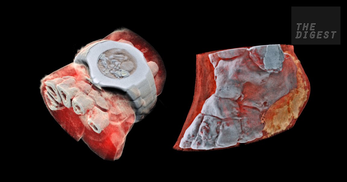
THE FAMILY BUSINESS. Phil and Anthony Butler aren’t just father and son. The physics professor and bioengineering professor (respectively) are also business partners. And this week, their company, MARS Bioimaging, unveiled a first-of-its-kind x-ray scanner 10 years in the making.
First, a quick recap of how x-ray imaging works. When x-rays travel through your body, they’re absorbed by denser materials (bones) and pass right through softer ones (muscles and other tissues). The x-rays that pass through unimpeded hit a film on the opposite side of your body. These show up as areas of solid black. The places where the x-rays couldn’t pass through appear solid white.

IN LIVING COLOR. Back to the Butlers’ invention. Their scanner uses a combination of Medipix technology — tech first developed to help researchers at the European Organization for Nuclear Research (CERN) track particles using the Large Hadron Collider — and computer algorithms to produce colorful, 3D x-rays.
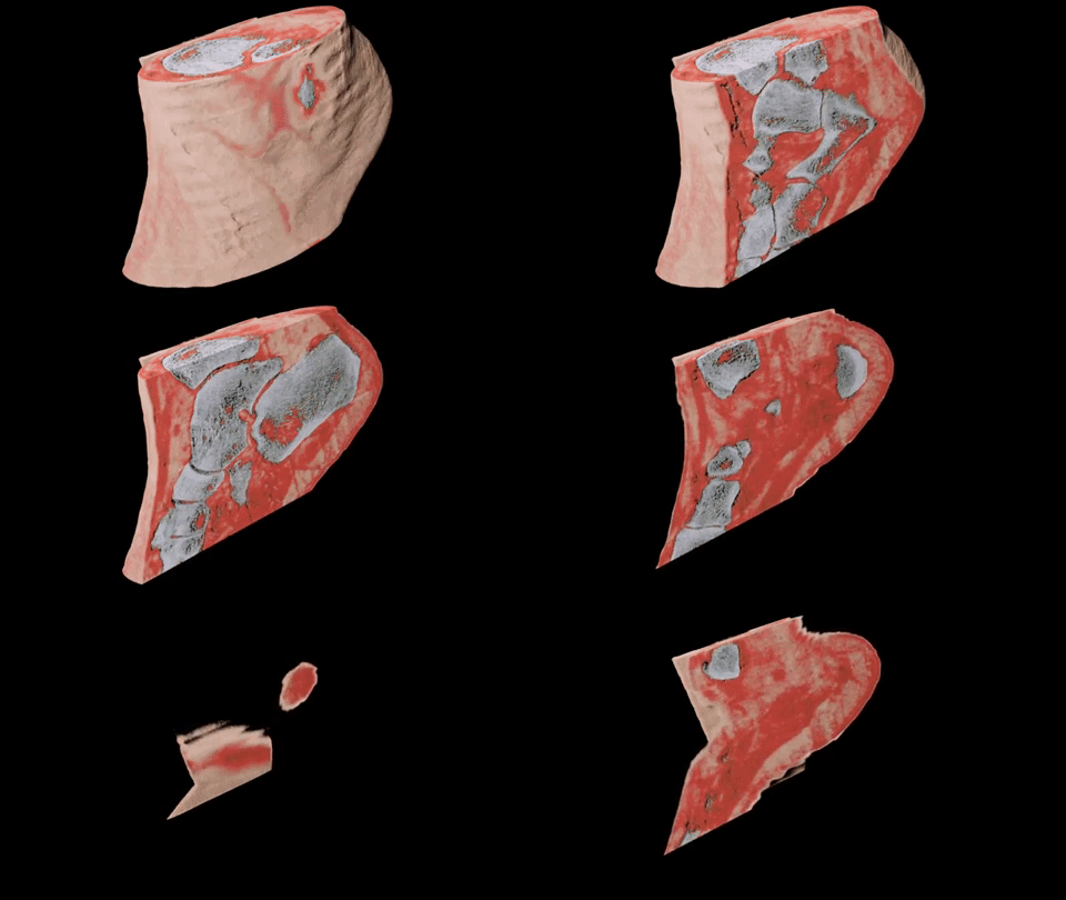
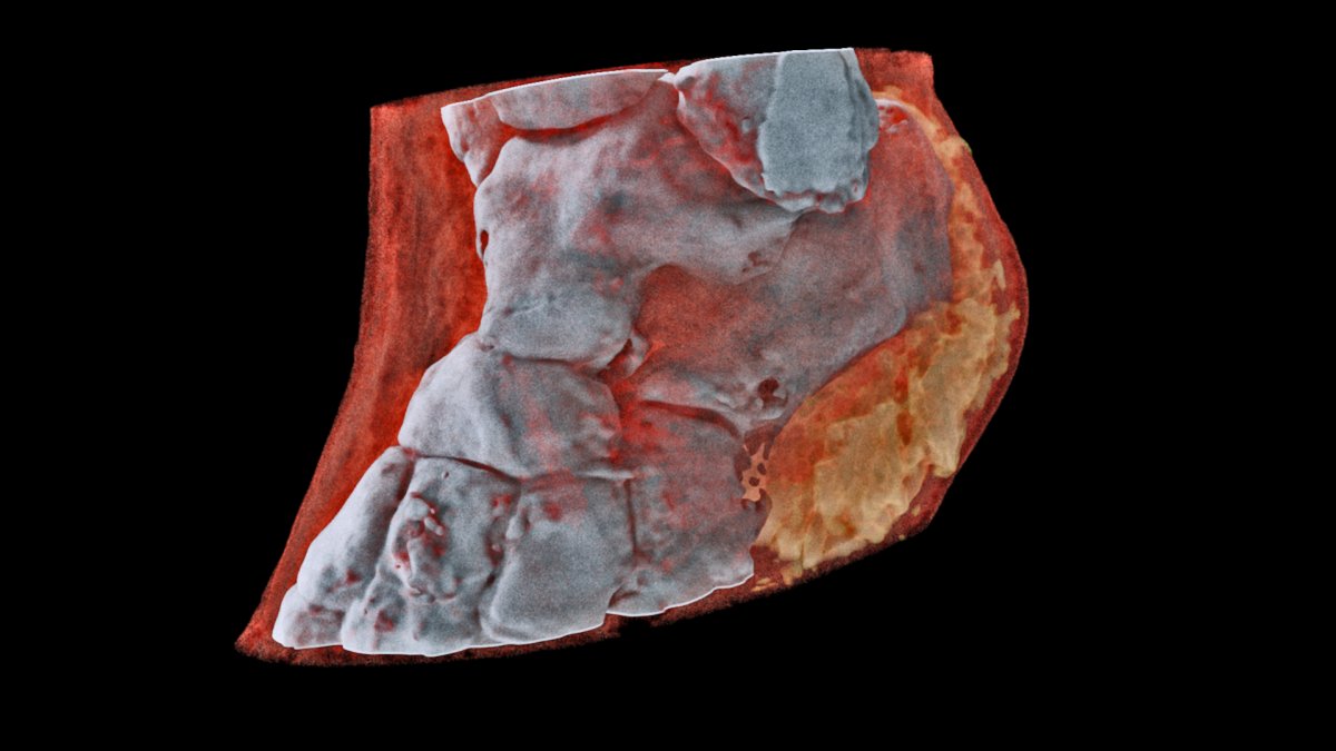
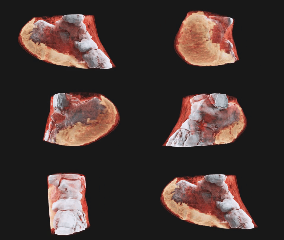
Instead of recording the x-rays as either passing right through the body or getting absorbed by the bone, this scanner records the precise energy levels of the x-rays as they hit each particle in your body. It then translates those measurements into different colors representing your bones, muscles, and other tissues.

BETTER DIAGNOSTICS. While the flat black-and-white x-rays doctors currently use are typically enough for them to notice if the bone in your arm has a fracture, they reveal very little about the tissue and muscle surrounding that bone. Doctors could use these new 3D x-rays to help diagnose issues in the bone and everything around it, too.
“This technology sets the machine apart diagnostically because its small pixels and accurate energy resolution mean that this new imaging tool is able to get images that no other imaging tool can achieve,” Phil Butler said in a CERN news release.

TESTING AND MORE TESTING. The MARS scanner is already in use for a number of studies, including some focused on cancer and strokes. As Anthony Butler told CERN, “In all of these studies, promising early results suggest that when spectral imaging is routinely used in clinics it will enable more accurate diagnosis and personalization of treatment.”
Next, the researchers plans to test out their scanner in a trial focused on orthopedic and rheumatology patients in New Zealand. Even if all goes well with that trial, however, it could still be years before the device secures the regulatory approval it would need for its use to become widespread.
READ MORE: First 3D Colour X-Ray of a Human Using CERN Technology [CERN]
More on x-rays: Watch out, Doc. An AI Is Going to Start Interpreting X-Rays