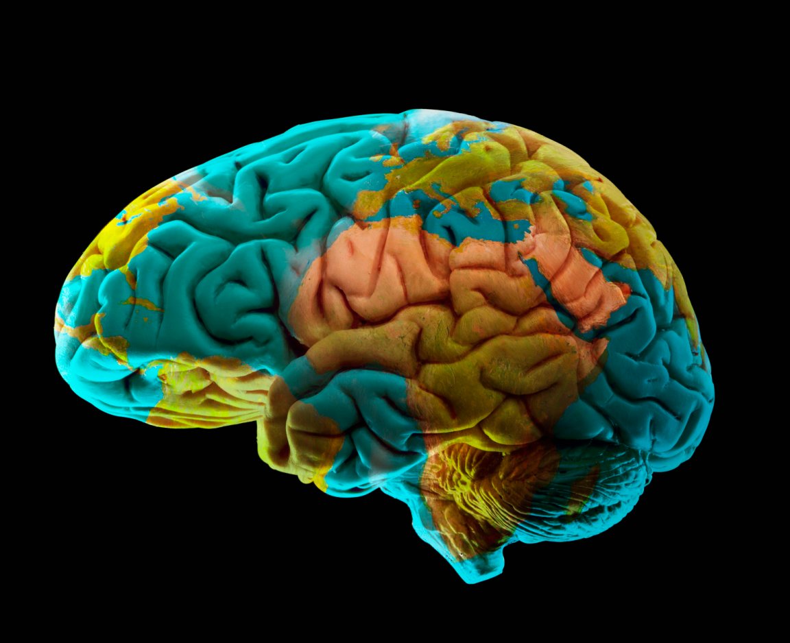
MAPPING THE BRAIN
Even with our growing knowledge of the cosmos, we are relatively clueless about how our own brains function. That’s why making neurological maps is such an important exercise — it allows us to see the structural basis of how our brains work.
The Allen Institute for Brain Science has just created one of the best maps ever. The Seattle-based organization published a comprehensive, high-resolution atlas of the entire human brain.
“This is the most structurally complete atlas to date and we hope it will serve as a new reference standard for the human brain across different disciplines,” said Ed Lein, investigator at the Allen Institute, in a press release.

The researchers put a donor brain through MRI and diffusion tensor imaging and then sliced it up by specific regions. The end result is a map of 862 annotated structures that comprise the human brain.
Studying the brain is so complex that researchers also had to create an entirely new scanner. The machine can image tissue sections the size of a complete human brain hemisphere at the resolution of roughly a hundredth the width of a human hair.
And in a bid to make the map a gold standard for brain research, the atlas was published both as a peer-reviewed paper in The Journal of Comparative Neurology and an online collection that anyone can access.
FILLING NEEDS
The new brain atlas fills a niche in a surprising vacuum of reliable brain maps. “Human brain atlases have long lagged behind atlases of the brain of worms, flies or mice, both in terms of spatial resolution and in terms of completeness,” Lein said.
But new technologies have thankfully spurred further development in this field. A similar map of the brain was published earlier this year by the Human Connectome Project. The main difference is the Allen study used only one brain, while the Human Connectome Project mapped 210 brains.