The deadly COVID-19 virus has killed more than a thousand over the past month, and scientists across the world are racing to understand the novel pathogen.
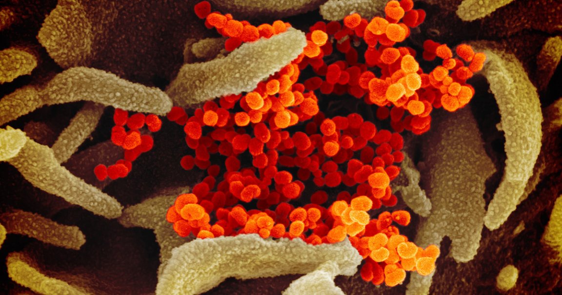
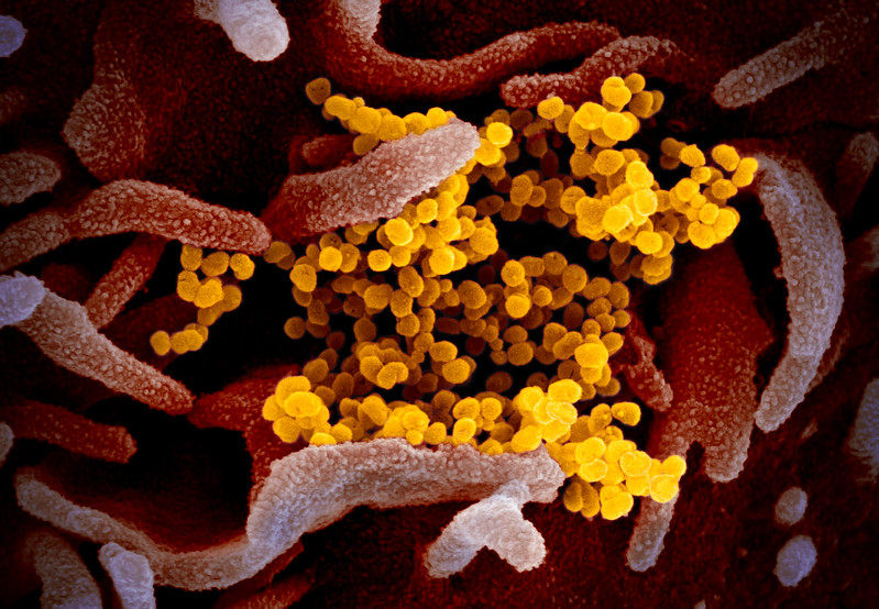
Now, researchers at the Montana-based National Institute of Allergy and Infectious Diseases’ Rocky Mountain Laboratories have released a gallery of stunning images of the virus that they took with a variety of scanning and transmission electron microscopes.
The haunting shots that remind you that your body is a microcosmic battleground of trillions of cells, many of them fighting off invaders that can range from the COVID-19 to the common cold.
Don’t believe us? A scanning electron microscope caught this cluster of the viruses nestled in the folds of a human cell from a patient in the US:

In this shot, from a transmission electron microscope, the viruses look almost like celestial objects:

Okay, one more. This scanning electron microscope shot shows the viruses living on the surface of cave-like cells cultured in a lab:
