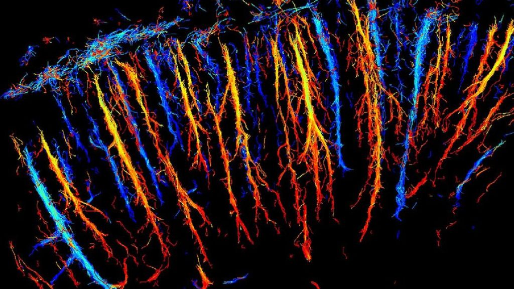
Ultrasound on a Live Rat Brain
Ultrasound is a type of imaging that uses high-frequency sound waves to look at organs and structures inside the body. Ultimately, this technique is best known for its use in relation to pregnant individuals, as it is the method that we use to check on the development and health of a fetus in utero; however, it’s also used to view the heart, blood vessels, kidneys, liver, and other organs.
Now, we can get views that are even smaller and more nuanced. Indeed, we can get microscopic images from it, and add ‘building a 3D view of blood vessel networks’ to its uses.
Researchers from France used ultrasounds to scan the brain of a live rat and rapidly construct a 3D view of a network of blood vessels in microscopic detail. For the procedure, the rat was injected with millions of very tiny bubbles, which reflect sound waves much better than blood vessels. As human soft tissue is nearly 90% water, ultrasound easily propagates in us. The bubbles augment the sound waves, allowing for a clearer end picture.
High-frame Rate Ultrasound
Besides the microbubbles, the primary reason why such a sharp and high-resolution image was able to be obtained comes thanks to scanning at a high frame-rate. Instead of taking an extended period of time acquiring a single, beautifully detailed image, the researchers took more than 500 images every second and then compared them.
They built a system that is able to compile those thousands of images and create a single, high-resolution view by looking at the differences among them that are caused by the bubble movements.
Within around 120 seconds, 75,000 images were collected and compiled to construct a 3D view of the rat’s brain with pixels just 0.01 mm in size. The team hopes that the system could reach the clinic and help with cancer and stroke diagnosis within a few years.
Clinical experiments have already begun on liver scans.