Hyperlensing Nano-vision
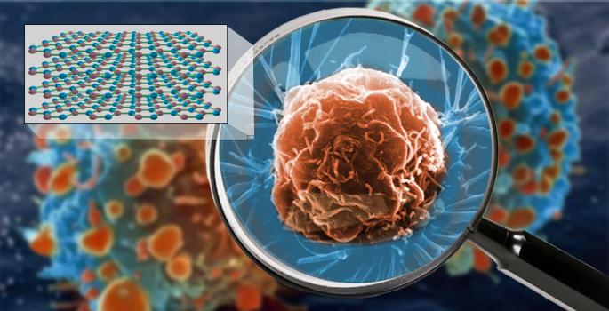
Research from Vanderbilt University and other participating institutions have devised a new lensing technology that allows scientists to see living cells in their natural environment. The new lens is so powerful that it can allow researchers to spot a small virus on the surface of a living cell. This remarkable resolution is thanks to advances in hyperlensing, a method of creating lenses with the ability to capture objects smaller than the wavelength of light.
In a press release from Vanderbilt, team member Alexander Giles, research physicist at the U.S. Naval Research Laboratory, said “Controlling and manipulating light at nanoscale dimensions is notoriously difficult and inefficient. Our work provides a new path forward for the next generation of materials and devices.” Prior to this development, it was possible to view objects in the nanoscale without hyperlensing, using technology such as electron-based and atomic-force microscopes. However, the application of these technologies was prohibitive as they could only operate under a high vacuum, they would bombard samples with harmful radiation, or could only be used with freeze-dried cells. Viewing living nanoscale objects in their natural environment is impossible using these techniques.

The laws of physics make it impossible for traditional lenses to resolve objects less than the wavelength of light. With infrared light, this barrier, known as the “diffraction limit,” doesn’t allow imaging to capture objects smaller than roughly 3,250 nanometers. The material devised by this research team is able to capture images of items as tiny as 30 nanometers in size. For perspective, the human hair has a diameter of 80,000 to 100,000 nanometers (nm).
Super Vision
The hyperlens is composed of hexagonal boron nitride (hBN), a naturally occurring crystal. Previous lenses devised with the material have been able to image objects as minuscule as the smallest known bacteria. The new research has led to an tenfold improvement in imaging capability, now equipping scientists with the ability to view many viruses, which can range in size from 20 nm to 400 nm.
Taking this ability to view small objects in tandem with the harmless nature of the imaging technique, and we can see that with hyperlensing researchers have acquired the ability to view cell processes in their natural environment, which will surely lead to significant advances in medical and biological science. The ability to view processes such as how a virus enters a cell could equip researchers with new avenues to fight them, or see how our immune system works on the cellular level could show us new ways of bolstering its capabilities. The researchers are confident that their findings via hyperlensing can even be improved with more research using larger crystals. “Currently, we have been testing very small flakes of purified hBN,” said Joshua Caldwell, associate professor of mechanical engineering at Vanderbilt University and research lead on the study. “We think that we will see even further improvements with larger crystals.”
The development of imaging has come a long way since the invention of the original microscope in the 17th century. We now have the ability to view the most intricate components of life. The impact this will have on biology and medicine is unknowable at this time, but the prospects are unlimited.