Getting Atoms to ‘Behave’
Students at the Leiden University’s Institute of Physics have developed a new nuclear magnetic microscope (NMR) that is 1,000 times more sensitive than current NMR microscopes. Even more, the microscope can function at temperatures close to absolute zero. The new microscope, which would aid in capturing 3D images in atomic resolution, uses the same technology as magnetic resonance imaging (MRI) machines.
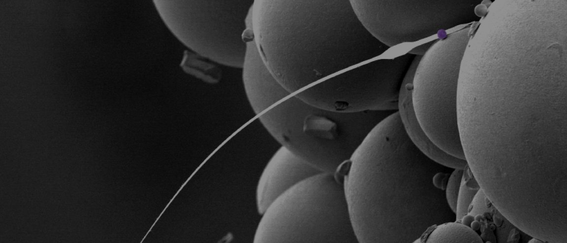
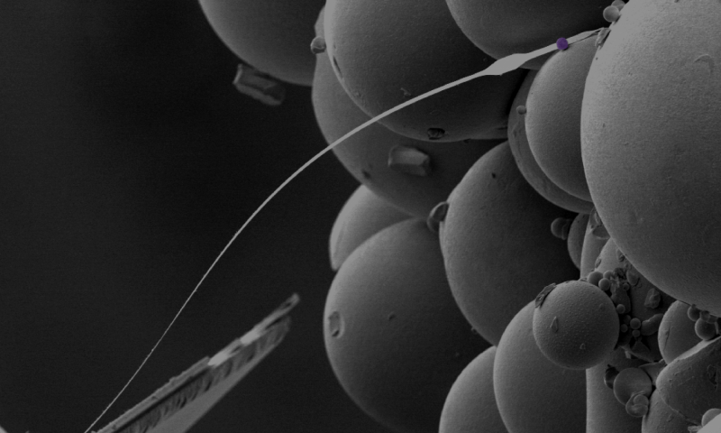
The revolutionary instrument uses a thin wire and a small magnetic ball that causes a consistent magnetic field which then provokes the surrounding atomic nuclei to line up in the same direction. Next, radio waves are sent through the sample, causing some nuclei to flip the other way—giving away their positions based on the time it took for them to flip back again and generating an image mapping their locations.

The ability of the microscope to work under extremely low temperatures presents another breakthrough in imaging atoms.
“The higher the temperature, the more the atoms will move a little bit,”says Jelmer Wagenaar, one of the PhD students who co-created the microscope. “It is like taking a photo — when somebody moves very fast, the photo gets a bit blurry. But the more that somebody sits still, the sharper the image. At zero Kelvin, the atoms do not move anymore, so the closer you work at zero Kelvin, the better it is to obtain sharp images.”
He adds that even at room temperature, 0.0001% of the nuclei’s spins align while the rest behave chaotically. “By decreasing the operating temperature to 42 miliKelvin, we achieved a three-order magnitude higher percentage of aligned spins,” he explains.
Medical ‘Holy Grail’
Just as NMRs made their way to hospitals in the form of MRIs, the technology is expected to open up a revolutionary window in the medical field.
“The holy grail here is 3D imaging biological tissues with atomic resolution,” Wagenaar says. “One example is that you might be able to use our technique to study Alzheimer’s patients’ brains at the molecular level, in order to find out how iron is locked up in proteins.”
He admits, however, that it will take time before the technology is developed well enough, but that this would lead to far more non-invasive imaging that would yield more accurate observations, such as images of proteins exactly the way they are folded—without damaging or disturbing them in the process.