
A Clear View
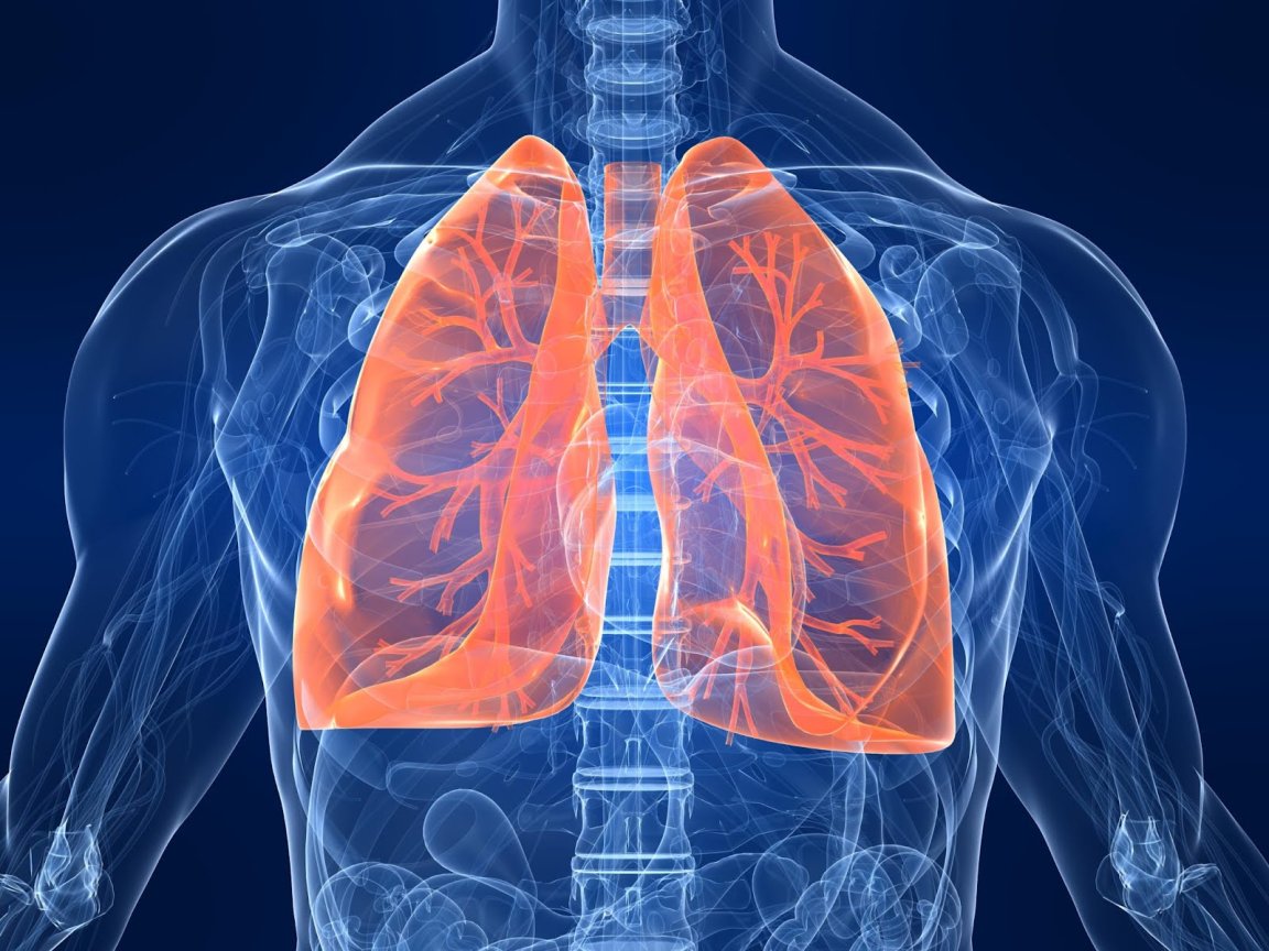
Traditional MRI scans use hydrogen protons in the body as molecular targets to give a picture of tissue. This works well for certain needs, but it doesn’t give a detailed picture of the lungs, as they’re full of air.
Oxygen is kind of important to our longevity, so there wasn’t really a way for doctors to combat this obstacle—until a new method was introduced.
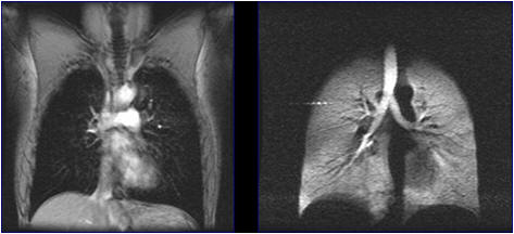
Scientists at the Sir Pete Mansfield Imaging Center have developed a new process that will give a clearer picture of lung disease using specially treated krypton gas. Once inhaled, the gas helps the spaces inside the lungs stand out in an MRI scan. This novel imaging method called “Inhaled Hyperpolarized Gas MRI,” uses lasers to “hyperpolarize” a noble gas, which then aligns the nuclei of the gas so it shows up on an MRI scan.
Controlled Explosions
The team from the the University of Nottingham announced that they’ve developed a new technique to generate hyperpolarized krypton gas at high purity. A higher purity means that the gas will work even better as a contrast agent for pulmonary MRI.
Chair in Translational Imaging at the Sir Peter Mansfield Imaging Centre, Professor Thomas Meersmann, said:
“To hyperpolarize the krypton-83 gas we diluted it in molecular hydrogen gas for the laser pumping process. After successful laser treatment the hydrogen gas is mixed with molecular oxygen and literally exploded it away in a safe and controlled fashion through a catalyzed combustion reaction.
“Remarkably, the hyperpolarized state of krypton-83 ‘survives’ the combustion event. Water vapor, the sole product of the ‘clean’ hydrogen reaction, is easily removed through condensation, leaving behind the purified laser-polarized krypton-83 gas.”
This development significantly improves the potential usefulness of laser-pumped krypton-83 as MRI contrast agent for clinical applications.
Hyperpolarized krypton-83 is currently being developed for whole body MRI at high magnetic field strength in the Sir Peter Mansfield Imaging Centre’s large 7 Tesla scanner. Studies will be carried out first on healthy volunteers before progressing to patient trials at a later phase.