Developing Human Connectome Project
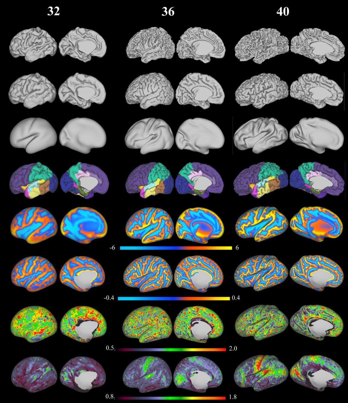
Scientists at the Developing Human Connectome Project (DHCP) are mapping the brains of babies in the hope that the images will help unravel the mysteries of neurological disorders. Thus far, the brains of 1,000 sleeping newborns have been mapped, as well as the brains of 500 babies that were still in utero. The researchers just released thousands of images of the brains of 40 of the newborns studied during the first phase of the project.

Because the high-resolution images require two to three hours of scanning, the process of obtaining them is tricky. When a sleeping newborn wakes up, all bets are off, as too much motion makes the scanning impossible. To improve their chances of success, the researchers adjusted the scanner software to ensure it wouldn’t make noise and could correct for slight movements computationally. Of course, even a baby sleeping soundly might wiggle or cry out, and even that minor motion can ruin an image.

Probing the Brain’s Subtle Wiring
The project’s ultimate goal is to create the first 3D map of the gestational human brain from 20 weeks onward. They will create the map using millions of images, one taken each second, which they will then stitch together into a coherent whole. Some of the babies in the study are identified as being at a high risk of developing autism and other neurological disorders. Researchers will ideally be able to create 3D maps of high risk and average risk brains for comparison.

In the longer term, the researchers hope to have both genetic and medical data for the babies, along with their test results as they grow older. This holistic, long-term view of brain development and health outcomes would afford scientists unprecedented insights into how even the most subtle changes in the anatomy and wiring of the brain might influence health and behavior later in life, perhaps leading to new avenues of treatment for various disorders.