
A Closer Look
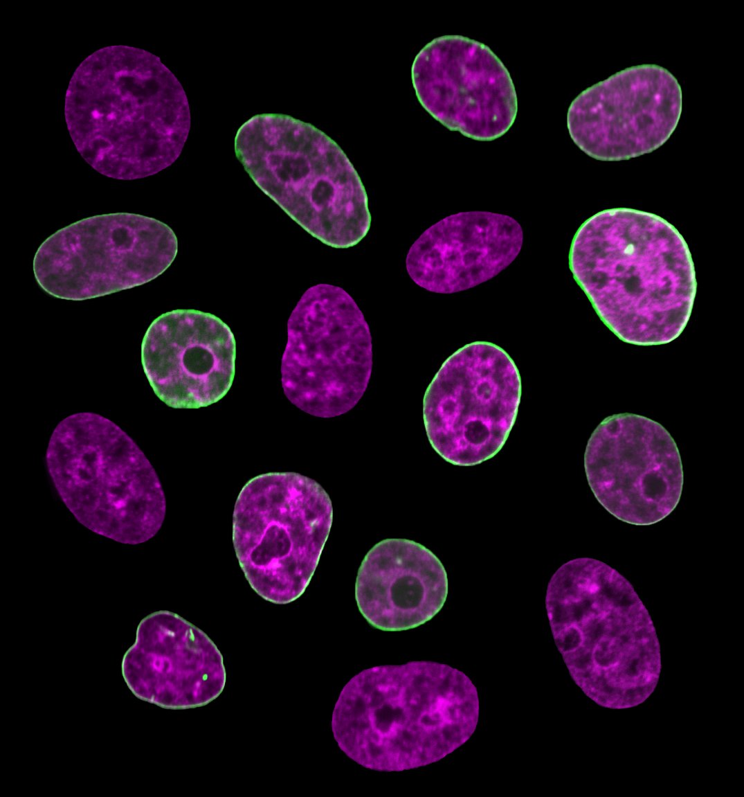
A new study by researchers at New York University (NYU), as published in the journal Proceedings of the National Academy of Sciences (PNAS), has established a method of gauging what stage of the cell cycle a living cell is currently at. Previously, it was only possible to take such measurements when working with a dead cell.
It was already known that the size and shape of a cell nucleus typically undergo big changes over the course of a cell’s lifespan. However, the challenge of taking measurements from a living cell meant that we didn’t know whether its shape changed over a shorter span of time.
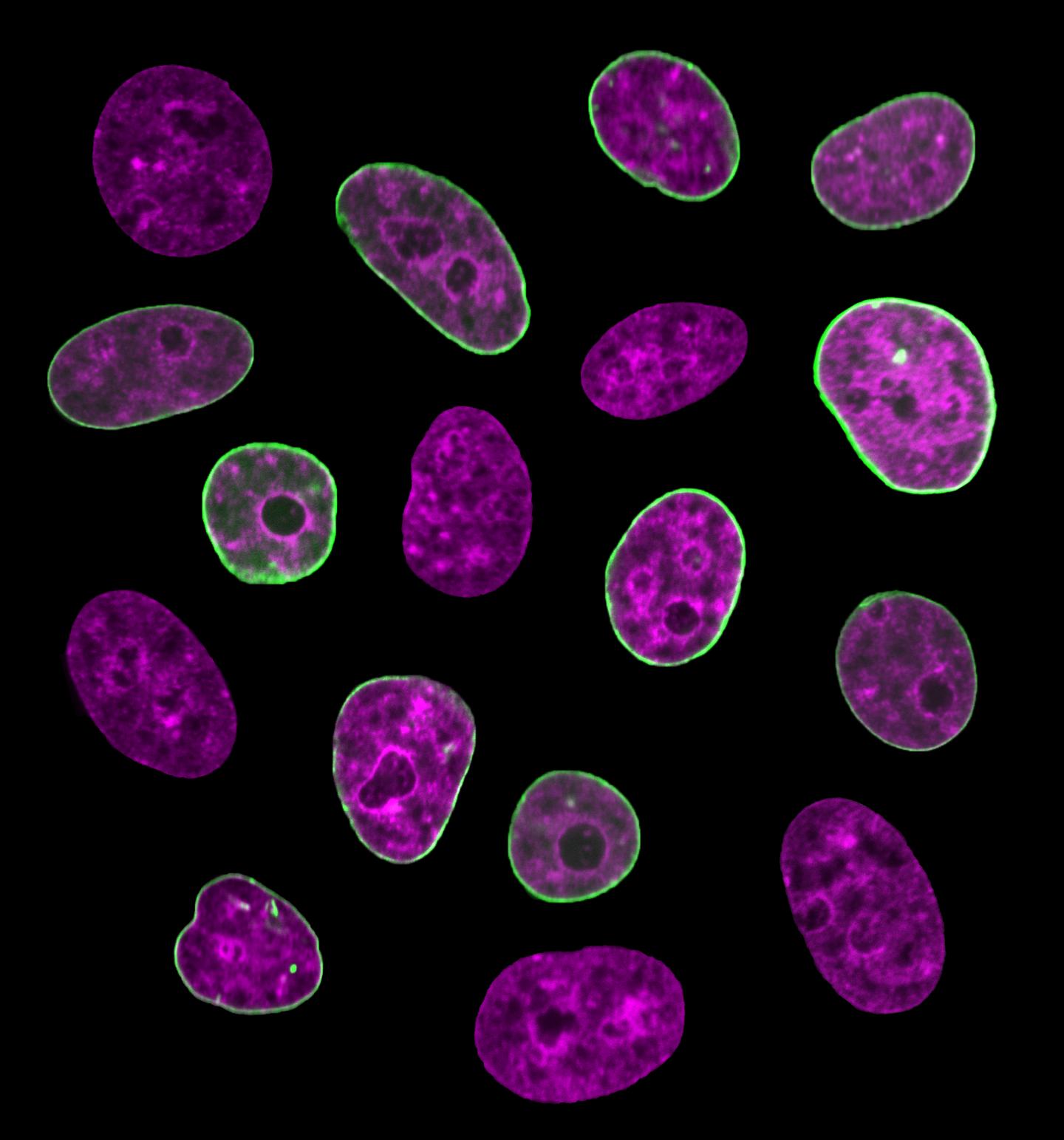
Using a cutting-edge fluorescence microscope, researchers were able to observe a previously undetected flicker of the nuclear envelope, which takes place over the course of just a few seconds. The amplitude of this fluctuation was seen to decrease as the cell cycle went on.
This motion could serve as an internal clock, as scientists could take measurements in order to understand what point in the cell cycle a living cell is currently occupying.
A Better Understanding
Being able to discern where a cell sits in its life cycle will hopefully facilitate a greater understanding of the most basic processes of human biology. This discovery stands to improve our knowledge of both healthy and diseased cells.
“We know that structural and functional errors of the nuclear envelope lead to a large number of developmental and inherited disorders, such as cardiomyopathy, muscular dystrophy, and cancer,” Alexandra Zidovska, the paper’s senior author and an assistant professor of physics at NYU, said in a statement released by the school. “Illuminating the mechanics of nuclear shape fluctuations might contribute to efforts to understand the nuclear envelope in health and disease.”