
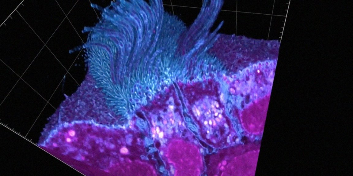
A stunning, ultra-detailed set of 3D images showing human respiratory cells infected by SARS-CoV-2, the virus that causes COVID-19, shines new light on how infections take hold, and how doctors might be able to battle them.
In order to actually see the cells and the tiny structures that coat their surfaces, the Utrecht University scientists had to quickly make some improvements to a microscopy technique called expansion microscopy, according to research that was shared online earlier this month but hasn’t yet been vetted for publication by an academic journal. Their updates allowed them to look at objects 20 nanometers wide in a microscope, New Scientist reports, a tenfold improvement that rendered the 100-nanometer-wide coronavirus clearly visible.
The images show multiple ways that the coronavirus alters and deforms infected cells as it multiplies, as highlighted in a video breakdown of the research produced by New Scientist.

Basically, the scientists discovered that the coronavirus, after infecting a host cell and multiplying as much as it can, digs outward from tiny filaments on a cell’s surface called microvilli with its spike protein. Knowing about this escape route — as well as the other ways that the images show the coronavirus deforming cells — could help scientists develop more effective drugs or treatments down the road. That’s speculative for now, of course, but as we continue to battle (read: get clobbered by) this pandemic, every little bit of new knowledge can make a huge difference.